Breast cancer can spread to the lungs or to the space between the lung and the chest wall making fluid build up around the lung.
Breast cancer chest wall invasion prognosis.
This can cause a build up of excess fluid a condition known as a.
This means that her2 positive breast cancer has a less favourable prognosis than her2 negative breast cancer.
Breast cancer cells can form in the region between the outside of the lungs and the chest wall also known as the pleural space.
Women younger than 35 years of age tend to be diagnosed with more aggressive higher grade tumours.
Early detection and diagnosis of breast cancer can significantly improve a person s outlook.
Breast cancer is staged from 0 to 4.
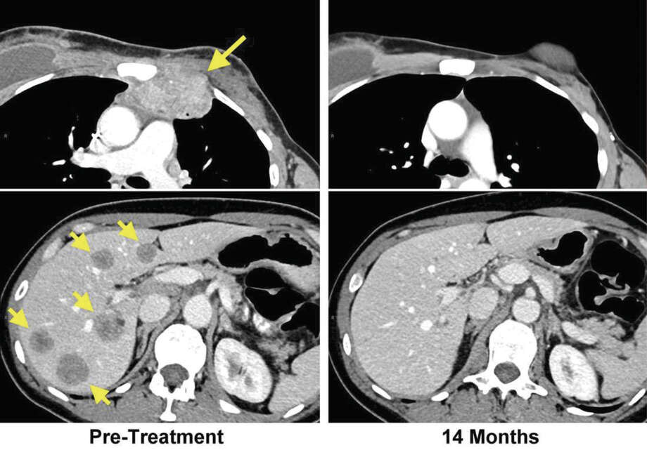
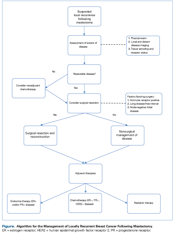
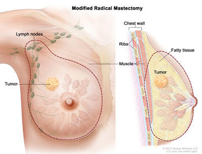
A chest wall recurrence is breast cancer that returns after a mastectomy a chest wall recurrence may involve skin muscle and fascia beneath the site of the original breast tumor as well as lymph nodes when cancer recurs in the chest wall it may be classed as a locoregional recurrence or it may be linked to distant metastasis if a chest wall recurrence is localized it is referred to as a.
The stage reflects tumor size lymph node involvement and how far cancer may have spread.
The relative five year survival rate for stage 3 breast cancer is 72 percent this means that out of 100 people with.
According to the acs the 5 year relative survival rate for localized breast cancer is 99.
Staging for breast cancer is very complex.
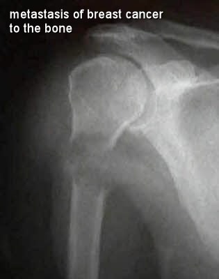
The physician will initially order an x ray to see if there is an abnormality.
Other things such as hormone receptor status and tumor grade are.
It is also called locally advanced breast cancer.
Their breast cancer is often more advanced at the time of diagnosis.
The tumor may be any size and cancer has invaded the chest wall or breast skin with evidence of swelling inflammation or ulcers such as with cases like inflammatory breast cancer the breast cancer may also have invaded up to 9 nearby lymph nodes.
Many different factors are considered before doctors can confirm your final stage.
If there is an abnormality shown on x ray your physician will then do a ct scan computed tomography or mri magnetic resonance imaging scan to gain additional information about the chest wall abnormality such as size and location.
Survival rates can be confusing and don t reflect your individual picture.


